


One life lost every 12 minutes.
This is the distressing reality of heart disease in Australia. One in four overall deaths are attributed to cardiovascular diseases (CVDs), to which men are 40% more likely to die than women. As you read this, three quarters of our population are in danger of developing heart disease, despite a large portion of risk factors being preventable simply through adequate diet, exercise, and weight loss. Heart Week, held in the first week of May every year, is a period dedicated to increasing awareness about CVDs, and to encourage people to undergo regular heart health check-ups to monitor and reduce their risks of developing them. Early diagnosis can save lives.
For you, as healthcare professionals, the safety and wellbeing of your patients is paramount. Countless hours of hard work and deliberation are poured into ensuring that you provide only the utmost quality of care, and diagnostic imaging. But all of this would be futile without regular checks in place to test your equipment for performance inhibiting faults that may prevent or delay an accurate diagnosis.
As the last day of Heart Week 2022 draws to a close, ProbeLogic would now like to take the opportunity to stress the importance of acoustically testing cardiac, and especially phased array probes. Throughout the week you have likely learnt sombre statistics, seen harrowing photos, and read touching anecdotes relating to the terrifying reality of cardiovascular disease in Australia. However, there is a significant element that you may not have been made aware of.
While we have been so desperately encouraging people to go get heart health check-ups to assess and diagnose their risk of developing CVDs, there’s a chance that the results that you have been returning are inaccurate. Yes, you read that right; the results that you yourself have agonised over producing, ensuring that every angle, measurement, and image is just perfect, so you have the most accurate information to aid a diagnosis. But how could this be possible? The answer lies not in an inherent flaw in your capabilities, but rather a fault in your equipment… and the scariest part is you may never know that your results are unusable.
Ultrasound techniques are widely used in the diagnosis of heart diseases. Cardiac sonography enables physicians to observe problems related to heart function; providing a clear view of veins and arteries. Doppler ultrasound is extremely effective as it can show the direction and speed of blood flow, assist in the detection of obstructions like blood clots and narrowed blood vessels, and help to diagnose regurgitation. In addition, duplex ultrasound being safe and devoid of radiation and contrast dyes has become the preferred diagnostic test.
Ultrasound probes, like the human heart, are made up of complex structures and internal mechanisms built to provide the best functionality for their user. Unfortunately, the complexity of the heart also results in a myriad of potential complications. So too can be said about your transducers which can experience a range of performance inhibiting faults. One such fault is downed or ‘dead’ elements which are incapable or producing or receiving an ultrasonic pulse.
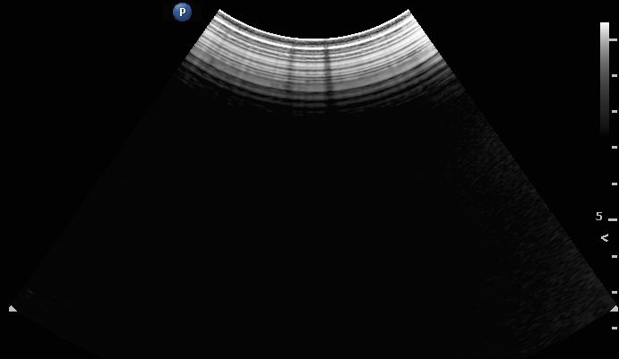
In regard to a standard linear probe, elements are fired sequentially to obtain slices or A-scan images which are then stitched together to create a complete B-mode image. When a few elements are down, it results in visible black lines in your image where no element is being fired. This is visible in the image shown above.
For phased array probes, elements are fired almost simultaneously, and rely on beamforming and interference patterns to construct an image. As these beams intersect, downed elements would not result in black lines, but rather a darker image which is then falsely compensated for by the operator who increases the image gain.
You may be wondering why a few downed elements matter in phased array probes if your image will appear relatively normal without them and can be compensated for by increasing the gain. To answer this question, we must first acknowledge that the darkness you will see is a direct result of less elements being fired. This may not seem significant for a B-mode image, but when we switch across to doppler (which often fires segments of 8 elements at a time), one or two missing elements makes a considerable difference to the recorded results.
This is especially true when interference in the doppler signal results in unwanted artefacts in the colour representation. While utilising pulsed wave or continuous wave doppler with downed elements, you may experience noise or obstructions in the measured signals which can return incorrect readings of velocity and thus result in misdiagnosis of regurgitation and other complications. Below is an example of a doppler study of the carotid artery – results at risk of being inaccurately measured due to downed elements.
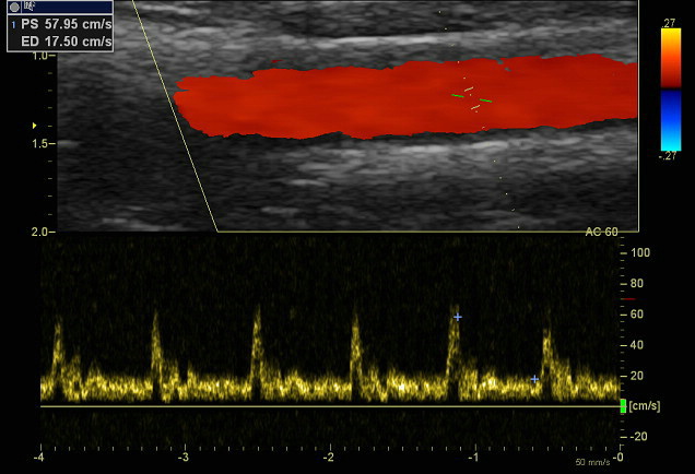
In a 2014 study, results confirmed that the degradation of an ultrasound probe through downed elements will negatively affect the quality of the Doppler-derived diagnostic information. The study also concluded that the results of Doppler measurements cannot be considered accurate or reliable if there are four or more contiguous dead elements in any given probe.
So how likely is it that your probe will experience these faults? Our experience has shown that approximately 25% of all probes on ultrasound systems aged 2 years or over, have some form of internal structural defect. Downed elements are an extremely common fault we see which can arise for various reasons including but not limited to impact related faults such as knocks or drops, wire damage due to cables being caught under machine wheels, damage to the array, and corrosion from fluid ingress. If caught early, most faults can be successfully repaired or mitigated against at a much cheaper cost than replacement.
This is why it is so important to have your probes routinely evaluated – especially phased array transducers used for cardiac applications. Acoustic testing can ensure that performance-inhibiting faults are identified before they reach your patients. ProbeLogic conducts comprehensive acoustic analysis and full transducer evaluation reports to provide quality assurance and peace of mind in your imaging. Our innovative, in-house designed StingArray tester provides data that you cannot obtain elsewhere. We recommend that probes are tested every 12 months, or immediately after impacts or noticeable damage. You can contact us for cost-effective ultrasound solutions including testing, repairs and consultations.
As professionals in the medical sector, your role as imaging providers is so significant in the diagnosis and subsequent treatment of cardiovascular diseases. Faulty probes have a proven effect on the quality of imaging, and thus pose a serious hazard to the accuracy of derived results. Testing is one clear way to reduce this risk. Remember, when your probes fail you, the results fail patients.
Are you willing to take the risk?